DR MILES DALBY
MBBS BSc MD FRCP FESC
CONDITIONS
Aortic Valve Disease
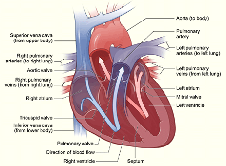
The heart has valves to ensure that the blood flow is in the right direction. The aortic valve is situated between the main pumping chamber of the heart (the left ventricle) and the main artery (aorta) at the ‘outflow’ of the heart. Whilst the heart is pumping blood out or ejecting (known as systole) the valve is open. When the left ventricle is relaxing and filling with new blood (known as diastole) the aortic valve is shut. The two main problems with the aortic valve are narrowing (Aortic Stenosis, AS) and leaking (Aortic Regurgitation, AR). Both have different effects on the heart however ultimately they can both cause serious problems with breathlessness and heart failure. If treatment to a heart valve is proposed it is preferable to do this before the heart becomes significantly damaged so early diagnosis and careful surveillance are important.
When investigating possible aortic valve disease in the clinic your cardiologist will need to take history and perform a physical examination. In addition they will want to perform some tests which will often include blood tests, a chest x-ray , ECG and echocardiogram. Further tests including a coronary angiogram and CT scan may also be necessary. Depending on the results your cardiologist may recommend continued follow up with medical therapy if appropriate and regular clinical surveillance including echocardiography. If intervention to the valve itself is needed this will usually require surgical aortic valve replacement (AVR) or transcatheter aortic valve replacement (TAVI).
Dr Dalby will discuss these tests, their implications and any subsequent tests, treatment or follow up with you so that you may reach a mutually agreeable management plan.